Existing Patients
(212) 679-4488
New Patients
(212) 401-2665

An echocardiogram (echo) is one of the first tests you receive to diagnose the cause of chest pain, shortness of breath, and other symptoms of heart disease. At Heartwise Cardiology, David Harnick, MD, and Raymonda Rastegar, MD, perform your echo in the office. This type of imaging produces fast, real-time results that reveal cardiovascular problems and allow quick, customized treatment. If you have questions about getting an echo or need to schedule an appointment, use the online booking feature or call the Murray Hill or Upper East Side office of Manhattan, New York today.
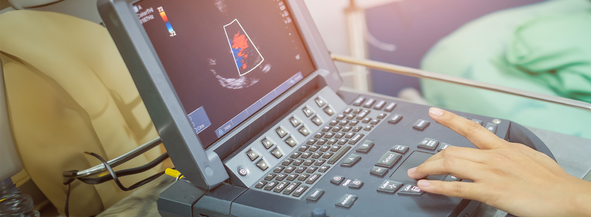
An echocardiogram uses safe, painless ultrasound (high-frequency sound waves) to produce images of your heart.
When you get an echocardiogram, your provider uses a handheld device called a transducer.
The transducer sends sound waves into your body. The waves bounce off tissues and return to the transducer. Then the device sends the information to a computer that produces real-time images of your heart and blood vessels.
Heartwise Cardiology performs an echo when symptoms like chest pain, dizziness, syncope (fainting), and shortness of breath suggest a heart problem. An echocardiogram helps them diagnose conditions such as:
Your provider also performs an echo to see how your heart is doing after treatment.
Echocardiograms reveal details about the heart’s structures. They can also show movement, such as muscle contractions, valve movement, and blood flow.
Your echocardiogram shows:
Your echo shows the health of your heart and how well it pumps blood.
Your provider may perform one of the following:
Transthoracic echocardiogram
This is the routine and most commonly used echocardiogram. After you relax on the exam table, your provider puts ultrasound jelly on the transducer or your skin and places the device against your chest. Then they move the device around your skin to get images of your heart.
You feel the transducer and slight pressure as your provider moves the device. However, an echo should never cause discomfort.
Transesophageal echocardiogram
This type of echocardiogram creates a sharper image of certain structures because the sound waves don't pass through your skin or ribs.
To ensure your comfort, your provider numbs your throat and gives you medication to help you relax. Then they gently guide a narrow transducer through your throat and into the esophagus.
Finally, your provider positions the probe in the esophagus where it passes near your heart and sends out the radio waves.
To learn more about echocardiograms or to schedule an appointment, call Heartwise Cardiology or book an appointment online today.
